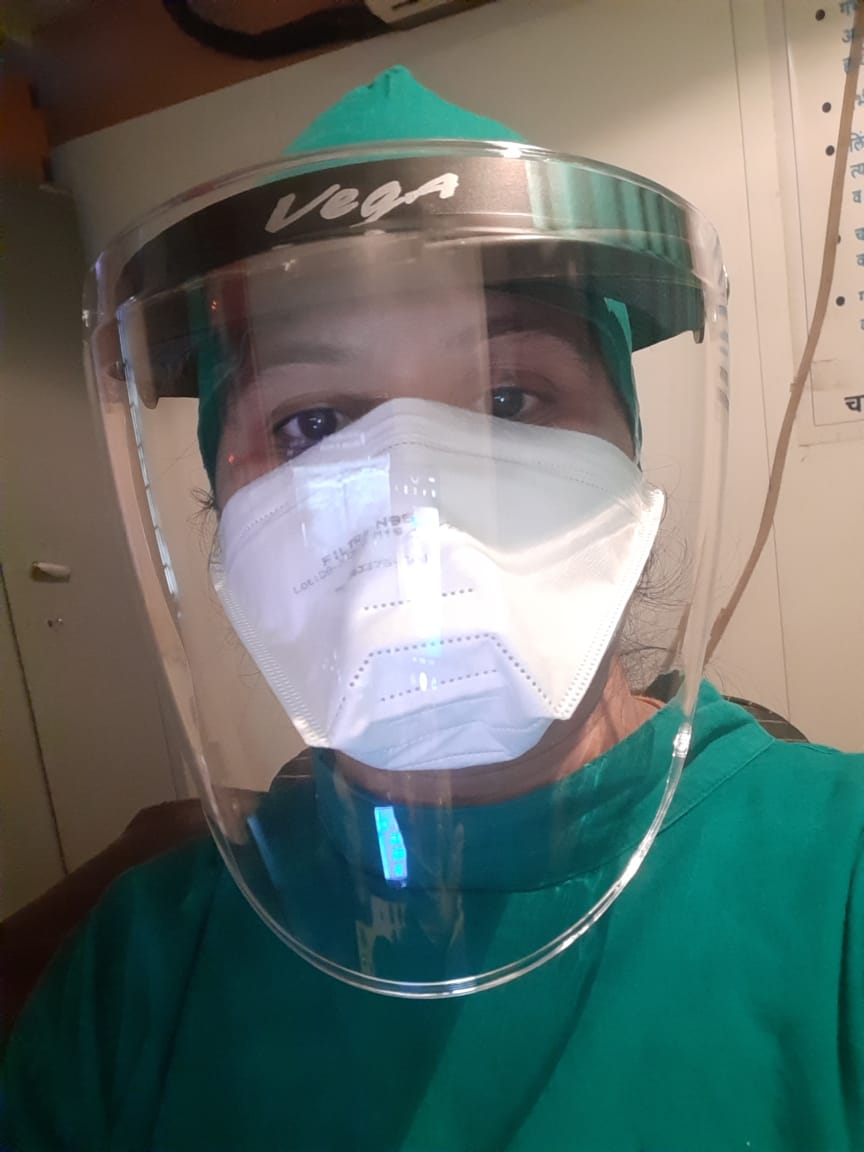
Dr. Manali Bafna, a medical graduate from S.R.T.R government medical college, Ambajogai, after completing her D.M.R.E (Radio-diagnosis) has hoards of experience in her specialty from various renowned institutions. She has over 9 years of experience as a practising Radiologist in diagnostic radiology.
Postgraduate from M.I.M.S.R medical college, Latur, she was under training for 2 years in DeenaNath Mangeshkar Hospital, Pune.
She further worked in radiology departments of government institutions like J.J. hospital (Mumbai) and C.P.R hospital (Kolhapur) as a Senior Registrar for considerable time.
While working as a consultant Radiologist for 4 years at the prestigious institute like Narayana Hrudayalaya, she undertook to the cause of creating awareness about Breast and Cervical Cancer and overall women's health and wellbeing. She further got trained in Fetal imaging.
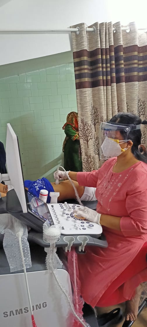
She excels at overall conventional and Sono-imaging including General, musculosketal, neurosonology, pediatric, fetal imaging, comprehensive women's imaging, all types of Colour Dopplers and Elastography.
She was responsible for infertility scans for patients undergoing IVF.
After relocating to Kolhapur, she was working with Apple hospital as a consultant Radiologist for 2 years.
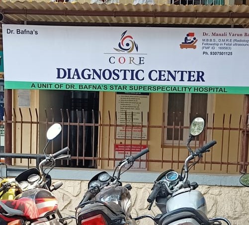
Now she has established her own diagnostic center, named CORE DIAGNOSTIC CENTER, with a sole purpose of providing Transparent and dependable diagnostic work up.
Core diagnostic center, which is a unit of Dr. Bafna's Star superpeciality hospital, works in basic conventional radiology (Digital x-ray) and Ultrasound.
Dr. Manali strives to provide the best medical services in the field of diagnostic radiology and also believes in providing high quality information and counseling to the patient regarding required further evaluation for their health condition and possible management options.
Explore this official profile to know more about the doctor, facilities and services to get credible health information, ask health questions, consult online or book an instant appointment with Dr. Manali V. Bafna.
PORTFOLIO
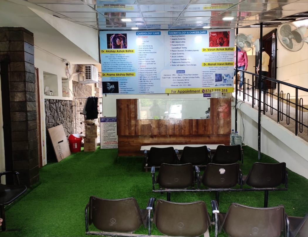
HOSPITAL INFO
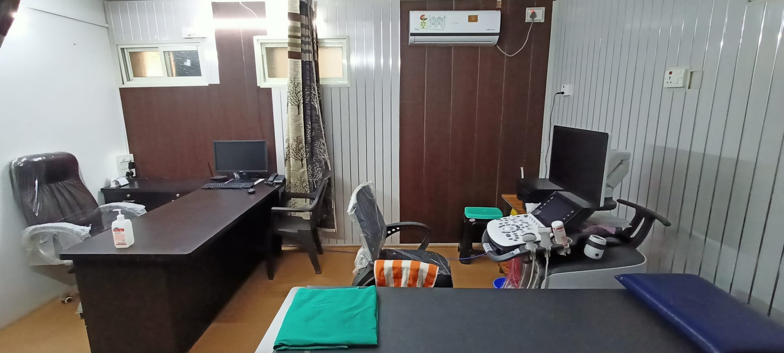
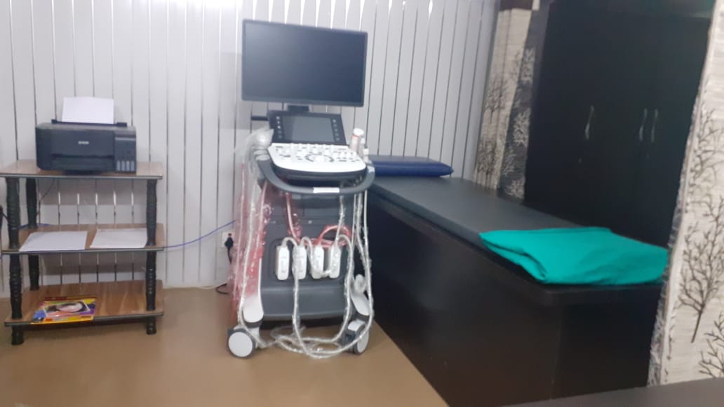
Core diagnostic center is Radiology unit catering to Ultrasound and Digital x-rays. We cover complete scope of diagnostic ultrasound including General, obstetric scan, Dopplers and all types of special scans.
The higher end machine ensures precision in the diagnostic work up. The diagnostic center's attachment to Star superspeciality hospital ensures emergency care and services in case of any medical emergency. The center is committed towards awareness in breast and cervical cancer imaging.
Paths to Radiology
Core Diagnostic Centre
Dr Bafna's Star Multispeciality Clinic & Hospital
OUR SERVICES
Color Doppler
Carotid
Scrotum
Renal
Portal
Venous
Arterial
Routine Ultrasonography
Whole Abdomen
KUB
Pelvis
Neck/ Thyroid
Musculo Skeletal
Orbit/ B Scan
Cranial/ Neurosonography
Breast/ Sonomammography
Follicular Monitoring
USG Chest
Obstetric Ultrasound
1st
Trimester
Dating Scan/ Fetal Viability
Scan
NT Scan (11 to 13.6 weeks)
2nd Trimester
Target Anomaly Scan / TIFFA (18 to 23 weeks)
Interval growth monitoring
NFT Scan (14 to 15.6 weeks)
Early Anomaly Scan (16 to 18 weeks)
Fetal Echocardiography
3rd Trimester
Growth Scan
Doppler/ IUGR Study
Near term scan for liquor/ weight
4D Scan (24 to 30 weeks)
AFI
Interventional Procedures
FNAC
Tissue Core Biopsy
Pleural/ Ascitic Fluid
Tapping
Digital X-ray
Doppler Ultrasound
Carotid Doppler
A physician prescribes a carotid ultrasound
for a variety of reasons, including if 1. You are a smoker and you have an
increased risk of having a stroke.
2. You have a blockage, known as an
occlusion, from plaque, a blood clot or something else.
3. your carotid artery is narrowing, known
as stenosis.
4. Your healthcare provider hears an
abnormal sound in your artery.
5. You had a TIA (transient ischemic
attack)
What
is a Doppler ultrasound?
A Doppler ultrasound is a special type of
ultrasound which shows blood flow as color images based on the speed and
direction of flow.
When
would you need a leg Doppler ultrasound?
1. When you have calf or leg pain
and swelling of the calf or ankle, then it might be suggestive of DVT. ~Alarm...!!!!!
2. Swollen vessels over leg and pain in leg. 3. To diagnose Superficial and
deep varicosities. 4. Bluish discoloration of toes.
Obstetric Doppler
Why might I
need a Obstetric Doppler scan? 1. You smoke 2. your baby isn't growing at a healthy
rate.
3. You've previously had a small baby
4. You've previously experienced a late
miscarriage or suffered the loss of your baby at birth. 5. You have a low or high BMI.
6. You're carrying twins or more.
7. You have an existing medical condition,
such as diabetes or high blood pressure.
NEUROSONOGRAPHY? WHEN?
1.
Demonstration or exclusion of an intracranial hemorrhage in a preterm neonate. 2. Follow-up of intraventricular hemorrhage
(IVH)-related complications to look for evolving findings pertaining to
ischemia. 3. Demonstration of ventriculomegaly,
congenital structural anomalies, intracranial vascular lesions. 4. Simple screening tool in the exclusion
of gross intracranial pathology.
Musculoskeletal Ultrasound
Musculoskeletal
Ultrasound ? When?
1. Tendon tears or tendinitis of
the rotator cuff in the shoulder, Achilles tendon in the ankle and many other
tendons throughout the body. 2. Muscle tears, masses or fluid
collections. 3. Ligament sprains or tears. 4. Inflammation or fluid
(effusions) within the bursae and joints. 5. Early changes of rheumatoid
arthritis. 6. Nerve entrapments such as
carpal tunnel syndrome. 7. Benign and malignant soft
tissue tumors.
8. Ganglion cysts. 9. Hernias. 10. Foreign bodies in the soft tissues
(such as splinters or glass).
11. Dislocations of the hip in
infants. 12. Fluid in a painful hip joint in
children. 13. Neck muscle abnormalities in
infants with torticollis (neck twisting). 14. Soft tissue masses
(lumps/bumps) in children.
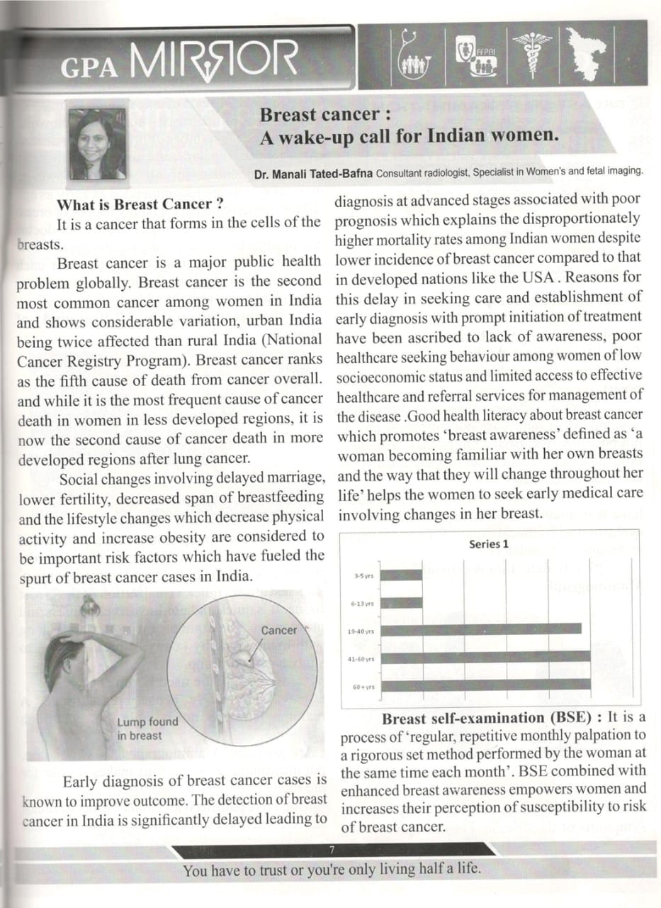
Breast ultrasound
Ultrasonography is effective and sensitive
in the diagnosis of breast cancer. It is also effective in diagnosing benign
breast diseases in younger women with dense breast tissue. Ultrasound is
cheaper and safer than other imaging modality for screening and diagnosis. Breast cancer when diagnosed early is
curable.
Ultrasound in pregnancy
Ideally, 5 periodic scans must be done in
pregnancy. A dating scan ( before 8-9 weeks), NT scan ( 11- 14 weeks), Anomaly scan ( 18- 20 weeks) Growth scans at 28 and 34 weeks.
Number of scans may increase/ decrease as
per condition of the pregnant woman/ opinion of treating doctor.
NT Scan
Why should
I go for NT Scan?
A nuchal translucency scan (also called
first trimester of pregnancy screening) is carried out during weeks 11–13 of a
pregnancy.
The scan uses ultrasound to screen for Down syndrome, or other
chromosomal or inherited conditions in the foetus.
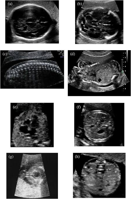
Anomaly Scan done
The mid-pregnancy anomaly scan is done for
checking any physical abnormalities in the growing baby. Although
it can’t pick up every problem, it gives the radiologist an idea about the
baby’s bones, heart, brain, spinal cord, face, kidneys and abdomen and allows
to identify the following conditions (some of which are very rare): • Anencephaly • Diaphragmatic hernia • Gastroschisis • Exomphalos • Open spina bifida • Bilateral renal agenesis • Lethal skeletal dysplasia • Edwards’ syndrome or T18 • Patau’s syndrome or T13 • Cleft lip • Serious cardiac abnormalities